In this post we learn Male reproductive system -organs and structure and external It is used in Biology, especially in Biology in Class XI Students studying in XI and XII are asked about Male reproductive system in the exam What is its external, what is its function and what are its parts and its function Give full information about this topic in the post I have given you but you should watch the post till the end Information about Male reproductive system Male reproductive system consists of parts for production of gametes and copulation . The male gonads are testes situated in scrotum , a pouch of pigmented skin arising from the lower abdominal wall. Its wall consists of smooth muscles called detox tunic muscles . It is divided into two compartments. -right and left by muscle septum . Each compartment encloses a testis , epididymis and a testicular end of a spermatic cord. It lies below the pubic symphysis in front of the upper part of the thigh and behind the penis.Scrotum protects testes and acts as thermo- regulator i.e. regulates temperature for proper functioning of testis . Testes are soft, smooth, pinkish oval organs about 4.5 cm long, 2.5 cm wide and 3 cm thick. Testes are mesodermal in origin and located outside the abdominal position . They are suspended in the scrotal sac by the spermatic cord . Each testis is connected to the wall of scrotum by a short fibromuscular band called gubernaculum . During early foetal life , the testes develop in the lumbar region of the abdominal cavity just below the kidney . During the seventh month of development , they descend permanently into the respective scrotal sacs through a passage called the inguinal canal. male reproductive system diagram

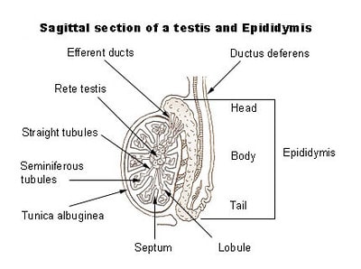
Male reproductive system organs In each testis, there are 200 to 300 lobules. In each lobule there are 1 to 4 convoluted loops called seminiferous tubules. These tubules converge towards the posterior surface and form a network of irregular tubules called rete testis. Testes descend into the scrotum along with peritoneum blood vessels and vas deferens. Histology of Testis : Testis is externally covered by fibrous connective tissue called tunica albuginea . It is covered internally by tunica vascularis formed by capillaries and externally by an incomplete peritoneal covering called tunica vaginalis . Seminiferous tubules : Are lined by cuboidal germinal epithelial cells. The germ cells undergo the process of spermatogenesis. Transverse section of the testis shows different stages of spermatogenesis like primary and secondary spermatocytes , spermatids and sperms. Few large pyramidal cells present interrupted between germinal epithelium are Sertoli cells. Sperm bundles get attached to Sertoli cells with their heads. Sertoli cells provide nourishment to the sperms till maturation .Between seminiferous tubules are present a few groups of cells called interstitial cells or cell of Leyding . These cells secrete the hormone testosterone after puberty Vasa efferentia: From the rete testis , 15 to 20 fine convoluted ductules , vasa efferentia , pierce the tunica albuginea to enter the caput or head of the epididymis. Epididymis : These are a pair of “c” shaped structures lying along the posterior border of each testis. Each shows presences of highly coiled duct, about 6 meters long, which is differentiated in three regions :- Upper wider head or output epididymis that receives vasa efferentia Middle narrower body or corpus epididymis. Lower duct is also wider, called tail or cauda epididymis. Male reproductive system -organs and structure In the head of epididymis , the sperms undergo physiological maturation acquiring increased motility and fertilizing capacity. In the tail , sperms are stored for a short period and then enter the vas deferens. Spermatozoa are produced irrespective of whether ejaculation takes place or not. The spermatozoa not ejaculate are reabsorbed in the vas deferens. Spermatozoa are produced irrespective of whether ejaculation takes place or not. The spermatozoa not ejaculated are reabsorbed in the vas deferens. Vasa deferentia : These are a pair of tubular structures arising from cauda epididymis . Each vas defences are about 40 cm long and enter into the abdominal cavity through the inguinal canal. It ascends in the form of spermatic cord, medically, towards the posterior wall of the urinary bladder Here it is joined by the duct from the seminal vesicle to form the ejaculatory duct. Ejaculatory duct : These are a pair of ducts each about 2 cm long . It is formed by joining of vas defences and a duct of seminal vesicles. Both ejaculatory ducts open into the urethra in the region of the prostate gland . They carry seminal fluid and spermatozoa to the urethra. Urethra
The male urethra provides a common pathway for the flow of urine and secretion of male reproductive organs called semen. The urethra includes three parts. The first is surrounded by the prostate gland and is called the prostatic urethra which carries urine only. The second part is the membranous urethra, which is situated between the end of the prostate gland and the root of penis. It carries both urine and semen. The third part is penile urethra which is situated in the penis. It carries both urine and semen . The urethra has two sphincters: an internal sphincter of smooth muscle fibers at its beginning , and external sphincter of striated muscles. Penis : It is cylindrical , erectile and pendulous organs suspended in pubic region in front of scrotum . Urethra passes through the length of penis . It contains three columns of erectile tissues. Ordinarily it remains small and limp but on sexual arousal , it becomes long, hard and erect , Erectile tissue has abundant blood sinuses . Blood flows in the sinuses and makes penis erect. The penis contains two postero- lateral tissues called corpora cavernosa and a median corpus spongiosum. Urethra passes through corpus spongiosum . Hence, It is also called spongy urethra. Near the tip of the penis , the corpus spongiosum is enlarged to form a soft and highly sensitive glans penis . It is covered by a loose retractable fold of skin called prepuce or foreskin. Male reproductive system -organs and structure Penis functions as a copulatory organ Male reproductive system structure Seminal vesicles : The seminal vesicles are two small fibro- muscular pouches present on the posterior side of the urinary bladder. Seminal duct joins with vas deferens and forms ejaculatory duct. Seminal vesicles secrete a viscous fluid fructose , fibrinogen and prostaglandins stimulate contractions in the female reproductive stimulate contractions in the female reproductive tract to help the process of fertilization . The fibrinogen helps the process of fertilization . The fibrinogen helps in coagulation of semen after ejaculation.
 |
| Image :- T.S of testis |
Prostate gland :- It consists of 20 to 30 separate lobes which open separately into the urethra. The prostatic fluid is a whitish liquid forming about 30 % of the total volume of semen. The prostatic secretion neutralizes the acidity of vaginal secretion . At PH 6.0 to 6.5 , sperms become motile and facilitate the process of fertilization . Cowper’s glands :- These are also known as bulbo-urethra glands. They are pea- sized and situated on either side of membranous urethra . These glands secrete an alkaline viscous fluid. Which neutralizes acids that may be present in the penile urethra due to previous urination and also lubricates the vagina of the female genital tract .
READ RELATED POST
- what is immune system
So far in this post you Male reproductive system - organs and structure , and their function
If you have any questions and suggestion in this post, so please comment and share with your friends