female reproductive system photos
In this post we learn
Female reproductive system - organs and structure and external It is used in Biology, especially in Biology in Class XI Students studying in XI and XII are asked about Female reproductive system in the exam What is its external, what is its function and what are its parts and its function Give full information about this topic in the post I have given you but you should watch the post till the end
female reproductive system photos
female reproductive system diagram

Image :- Female reproductive system
Female reproductive system - organs and structure
Female reproductive system organs
Labia majora : These are two large folds which form the boundary of the vulva. They are homologous to the scrotum of males. They are composed of skin , fibrous tissue and fat. These are prominent and longitudinal folds on right and left sides of the vestibule .
Labia minora : These are smaller and thinner lip-like folds located just medically to the labia majora . Posteriorly the labia minora are fused together to form the fourchette.
Mons pubis: It is fleshy elevation above the labia majora.
Clitoris : It is small erectile organs lying at the anterior end of the labia minora . It is homologous to the penis of males . It shows the presence of erectile tissues .
Vestibule : It is a median vertical depression of vulva enclosing vagina and urethral opening .
Hymen : It is a thin of mucous membrane which partially occludes the opening of the vagina
Vestibular glands : These are a pair of vestibular glands or Bartholin’s glands which occur on each side of the vaginal opening . These glands are homologous to the Cowper’s glands of the male. They secrete a lubricating fluid.
female reproductive system real photos
Breasis : These are a pair of rounded structures found in the pectoral region on the ventral thoracic wall . It has an erectile nipple in the middle. These are a pair of modified sweat glands . Each breast contains fatty connective tissue and numerous lactiferous glands. It has 15-20 openings of lactiferous ducts which carry milk from mammary glands to nipples . Lactiferous ducts dilate and from lactiferous sinuses just beneath the nipple to store the milk The base of the niple shows a dark brown rounded area called areola.
female reproductive system functions
Ovaries : A pair of ovaries are the primary sex organs of the female reproductive system . They are almonds . shaped bodies each measuring about 3 cm long , 1.5 cm wide and 1 cm thick . The ovaries lie in the lower part of the abdomen . Each ovary is suspended from the dorsal body wall (to broad ligament) by a fold of peritoneum . The mesovarium Ovary is connected to uterus by an ovarian ligament and is connected to uterus by an ovarian ligaments and is connected to laterals body wall by a suspensory ligaments
Ovaries produce ova and also female sex hormones - oestrogen and progesterone . These two hormones control menstrual cycle and secondary sexual characters
Structure of Ovary
Each ovary is a compact structure consisting of inner medulla and outer cortex. The medulla contains connective tissue called stroma. The cortex is lined by germinal epithelium . Oogonia arise from endoderm of the yolk sac and migrate to ovaries during embryonic developments
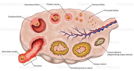
Image : Ovary
Female reproductive system - organs and structure
The process of Oogenesis begins even before the birth of a female baby . The ovaries contain 2 millions or more oogonia which become primary oocytes about six months before a human female is born . At the time of birth, about 1 million primordial follicles are present in each ovary and only about 40,000 remain by the time of puberty , the rest being degenerated . Out of these , during every menstrual cycle only one Graafian follicle reaches maturity and then ovulation takes place. The remaining follicles degenerate (atrophied ) Merch is at the age of about thirteen years and menopause (end of menstrual cycle ) is at the age of about forty five years . So the reproductive span is approximately thirty two years . There are about 13 menstrual cycles per year .
female reproductive system real photos
Histology of ovary :-
Tunica albuginea is a whitish capsule of dense irregular connective tissue located immediately inside the germinal epithelium.
Ovary shows cyclic changes during the menstrual cycle. Cortical region shows different stages of development of ovarian follicles or Graafian follicles . Each follicle contains a large ovum surrounded by many layers of follicle cells. The follicle cells of a maturing follicle secrete oestrogen.
The outermost granulosa cells rest on a basement membrane . Encircling the basement membrane is a region called theca folliculi . many capillaries are present in the theca folliculi
i) Theca interna - A highly vascularised internal layer of secretory cells.
ii) Theca externa - an outer layer of connective tissue cells.
Fallopian tubes (oviducts ) :- These are a pair of tubes lying horizontally over the peritoneal cavity close to the ovary . Each fallopian tube is about 10 to 12 pm long , narrow, muscular structure lined by ciliated epithelium . It conducts egg or ovum
discharge from the ovary to the uterus . It is supported by a double fold of peritoneum called mesosalpinx .
Female reproductive system - organs and structure
The free proximal end is dilated into a funnel-like infundibulum with a number of finger-like processes called fimbriae as its free borer. It shows the presence of an opening called ostium . This funnel is quite close to the ovary of its side so that the ova discharge from the ovary is received . Amulla is the site of fertilization . Cornua / isthmus is a very short , narrow part opening in the uterus .
Vagina - This is a highly collapsible and highly distensible fibro-muscular tube in which the cervix opens. It measures about 7.5 cm in length . It opens into the vestibule of vulva . inner lining cells of vagina store glycogen . Vaginal bacteria mainly Lactobacillus ferment the glycogen and this makes the mucus acidic.
READ RELATED POST
So far in this post you Female reproductive system - organs and structure , and their function
If you have any questions and suggestion in this post, so please comment and
share with your friends
female reproductive system photos
Next post
you can download this post
click download button